
Digital Reconstruction Reveals the Face
Goldlaner.com – For decades the skull of “Little Foot” — an Australopithecus individual who lived roughly 3.67 million years ago — resisted every attempt to examine its facial anatomy. Now a team led by Dr. Amélie Beaudet at the University of the Witwatersrand has completed a digital reconstruction that reveals the face in detail for the first time, exposing orbital architecture, nasal passages, and feeding structures that had been invisible since the specimen was buried in South African cave sediment.
The study, published in Comptes Rendus Palevol, marks a turning point for researchers studying one of the most complete early hominin skeletons ever recovered. Beaudet, an honorary researcher in the School of Geography, Archaeology and Environmental Studies in Johannesburg, spent years working with the fossil before the computational approach made the facial reconstruction possible.
How Four Tiny Bones Became a Landmark Skeleton
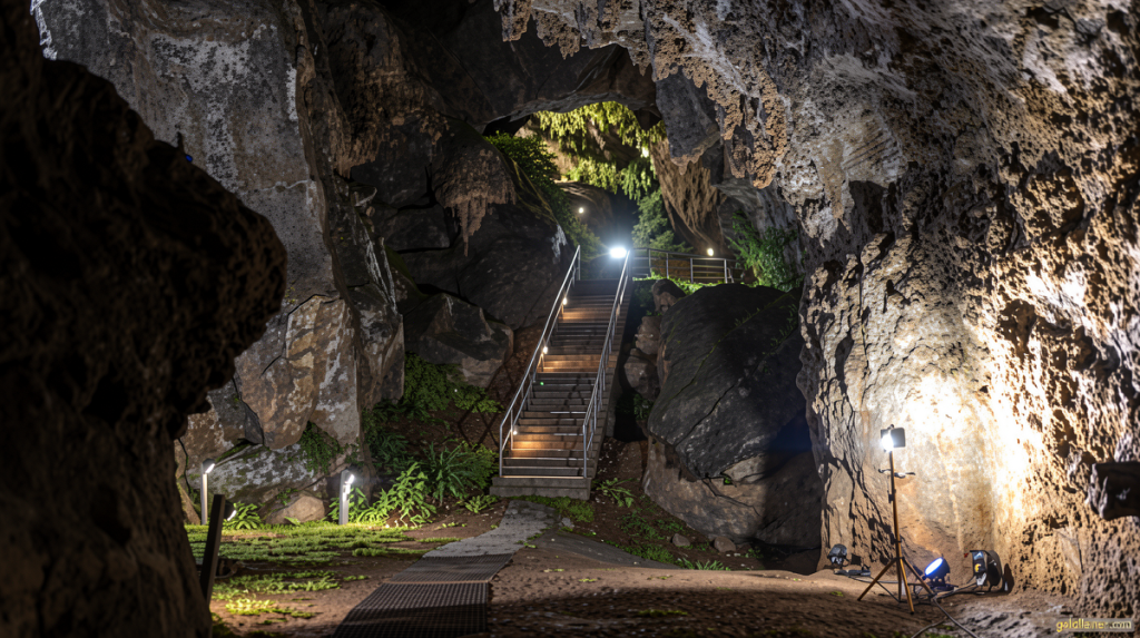
The discovery traces back to the Witwatersrand museum collection, where paleoanthropologist Ronald Clarke spotted four minute bone fragments that matched no known specimen. That observation sent him into the Sterkfontein Caves, northwest of Johannesburg, where during the 1990s he began extracting what would become the most complete Australopithecus skeleton known to science — approximately 90 percent intact.
The excavation consumed roughly two decades of painstaking work, freeing each element from a matrix of calcite-encrusted sediment. The resulting specimen, a chimpanzee-sized biped that retained powerful upper limbs for climbing, anchors southern Africa’s role in the earliest chapters of hominin evolution. Beaudet has stressed that the skeleton constitutes the oldest direct evidence of human evolution on the continent.
Why the Skull Demanded a Computational Solution
Millions of years of shifting sediment above and around the burial site had compressed and distorted the cranium beyond the reach of manual repositioning. Physical reconstruction would have destroyed the very evidence the bones carried. Instead, Beaudet and collaborators used virtual realignment to undo the geological deformation in software, restoring the facial elements to their anatomically correct orientations.
To capture internal detail at unprecedented resolution, the fossilized skull made its first journey outside South Africa. It was transported to the Diamond Light Source synchrotron at the Harwell Science and Innovation Campus in Oxfordshire, England, where intense, nondestructive X-rays produced more than 9,000 high-resolution images and terabytes of raw data. Those datasets were processed on a supercomputer at the University of Cambridge, which rendered the facial bones in three dimensions and completed the digital reconstruction of the face.
“Only a handful of Australopithecus fossils preserve an almost complete face, making Little Foot a rare and valuable reference point,” Beaudet said. “Little Foot’s face preserves key anatomical regions involved in vision, breathing and feeding, and its skull will offer further key elements for understanding our evolutionary history.”
Comparisons Across Africa and the Great Apes
Once the facial morphology was restored, the team compared it against three other Australopithecus specimens — one from South Africa and two from Ethiopia — as well as against modern great apes. The results were unexpected: Little Foot’s face size fell between that of a gorilla and an orangutan, while overall shape tracked more closely with orangutans and bonobos. Eye-socket dimensions and facial proportions aligned more closely with East African Australopithecus material, hinting at connections across the continent that had not been apparent from postcranial anatomy alone.
Frequently Asked Questions
Where was Little Foot discovered? The skeleton was excavated from the Sterkfontein Caves, part of the Cradle of Humankind World Heritage Site in South Africa, beginning in the 1990s under the direction of Ronald Clarke.
How complete is the specimen? Approximately 90 percent of the skeleton was recovered, making it the most complete Australopithecus skeleton known to science.
Why couldn’t researchers simply reassemble the skull by hand? Geological compression over millions of years had deformed the cranial bones to the point that physical repositioning would have destroyed the fossil evidence. Computational methods were required to restore anatomical orientation without altering the original material.
What imaging facility was used? The skull was scanned at the Diamond Light Source synchrotron in Oxfordshire, England, generating over 9,000 high-resolution images that were subsequently processed on a supercomputer at the University of Cambridge.
When was the study published? The findings appeared in the journal Comptes Rendus Palevol, with Dr. Amélie Beaudet of the University of the Witwatersrand as lead author.